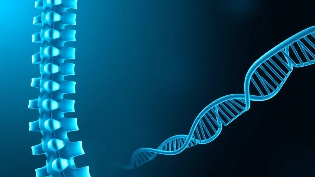
Researchers have uncovered how one of the most common causes of back pain takes hold — and, using fish, found hints that existing drugs might slow it.
Scientists at the University of Edinburgh and the University of Bristol studied intervertebral disc degeneration (IVDD), the wearing down of the cushions between spinal bones. Published August 6, 2026 in Communications Biology.
What an intervertebral disc is
Each disc has two parts, and their mechanical relationship is what makes the structure work.
A tough fibrous outer ring, built from concentric layers of collagen fibres oriented in alternating directions, surrounds a soft gel-like core. The core is largely water held by molecules that attract and retain it, and it behaves as an incompressible fluid.
Load applied to the spine compresses the core, which pushes outward against the ring, which contains it. That containment converts compression into distributed tension the ring is built to bear — a hydraulic arrangement that works well while the ring holds.
What goes wrong
The team found that disruptions to collagen IX, a protein that stabilises disc fibres, drive the disease.
Collagen IX is not the main structural collagen. It sits on the surface of the principal fibres and cross-links them to one another and to surrounding matrix, functioning as a connector that keeps the fibre network organised.
Losing it does not remove the fibres. It removes their organisation — and a fibre array that cannot distribute load as a unit fails progressively at whatever point bears most, which is a plausible account of how degeneration accumulates over years.
The downstream cascade
Along with that came changes in fat metabolism, the mTOR growth pathway, phosphate handling and vitamin A signalling, leading to mineral buildup that hardens the spine.
That combination is more informative than the structural defect alone. mTOR is a central regulator of cellular growth and nutrient sensing; phosphate handling directly governs whether tissue mineralises; vitamin A signalling controls cell differentiation, including whether cells adopt bone-forming behaviour.
Together they describe disc cells changing identity — behaving like bone-forming cells and depositing mineral in tissue that should stay flexible. That is a different disease process from mechanical wear, and it suggests intervention points that a purely structural account would not offer.
Why zebrafish
The team bred zebrafish lacking a working collagen IX gene. As the fish aged, their spines developed problems mirroring human disc disease — including fused vertebrae and abnormal mineralization.
Fish are an unobvious model for a spinal condition associated with upright posture and human load-bearing, and they earn their place for practical reasons. They develop rapidly, are transparent as larvae so skeletal development can be watched directly, and can be bred in numbers that permit drug screening at a scale mammals do not allow.
The fact that the same gene deletion produces recognisably similar pathology in fish also indicates the underlying biology is ancient and conserved — which strengthens the case that findings transfer.
Toward non-surgical options
A bisphosphonate — an existing osteoporosis drug — blocked the mineral buildup, and food restriction plus fat-metabolism-dampening drugs reduced spinal fusions.
Bisphosphonates work by binding to mineral surfaces in bone and inhibiting the cells that break bone down. Applying them here targets abnormal mineral deposition rather than bone loss — a different use of the same pharmacology, and one that follows directly from the phosphate-handling finding.
The dietary and metabolic result connects to the fat-metabolism changes, and it is the more speculative of the two: food restriction affects a great many pathways, so a benefit does not establish which one mattered.
Why repurposing matters here
“For decades, surgery has been the only real answer for disc disease,” said Erika Kague; the findings “point to several ways of slowing it down.”
Both interventions identified are already approved drugs, which shortens the path considerably. Safety in humans is established, manufacturing exists, and trials can begin without the years of preclinical toxicology a new molecule requires.
Why disc disease has resisted drug development
Back pain is among the leading causes of disability worldwide, and the near-total absence of disease-modifying drugs for it reflects specific obstacles rather than neglect.
The disc is the largest avascular structure in the body. It has no blood supply of its own, with nutrients diffusing in from adjacent vertebrae — which limits healing capacity and makes drug delivery genuinely difficult, since a systemically administered compound reaches disc tissue poorly.
Diagnosis is also imprecise. Imaging shows disc degeneration in large numbers of people with no pain at all, and some people with severe pain have unremarkable scans. The correlation between visible degeneration and symptoms is weak, which makes it hard to define who should be treated or to measure whether a drug helped.
Slow progression compounds the problem. A condition developing over decades requires long or well-chosen surrogate-endpoint trials, and no validated surrogate exists. Identifying a mineralisation process that can be measured on imaging would help with exactly that.
The work is early and in fish, but it flags concrete drug targets for a condition with few options. Research news, not medical advice.