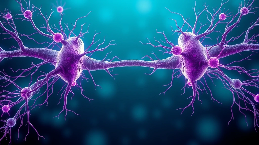
Scientists have used lab-grown human nerve cells to repair the breathing circuits damaged by spinal cord injury — at least in rats — an early proof-of-concept for a hard-to-treat problem.
Researchers at the Gladstone Institutes engineered human V2a interneurons from induced pluripotent stem cells and transplanted them into rats with neck-level spinal cord injuries. Published in Science Translational Medicine.
Why neck-level injury stops breathing
The diaphragm is controlled by nerves emerging from the spinal cord high in the neck, and those nerves receive their instructions from a breathing centre in the brainstem.
An injury between the brainstem and that level severs the connection. The diaphragm is intact and its motor nerves are intact, and the command signal can no longer reach them — which is why high cervical injury causes respiratory failure and long-term ventilator dependence, and why it is among the most consequential outcomes of spinal trauma.
The problem is a broken relay rather than destroyed machinery, which is what makes it a candidate for this approach.
What V2a interneurons do
These are nerve cells that relay signals within the spinal cord — not motor neurons commanding muscle, and not sensory neurons carrying information up.
V2a interneurons specifically participate in coordinating rhythmic motor output, including breathing, and they occupy an unusual position: they form alternative routes around the direct pathway. Some spontaneous recovery after partial spinal injury is attributed to these cells reorganising to carry signals through remaining tissue.
Choosing them is therefore a choice to amplify a repair mechanism the spinal cord already has, rather than to reconstruct the original circuit.
The engineering
The cells were designed to be activated by light and to be freezable for eventual clinical use — a process that took roughly 18 months to optimise.
Light activation is a research tool. Introducing a light-sensitive protein lets researchers switch the transplanted cells on at will, which is how they established the cells were functionally integrated rather than merely present and alive.
Freezability is the translational element and is easy to underrate. A therapy requiring cells to be made fresh for each patient is difficult to deploy — production must be scheduled against surgery, and any delay wastes the batch. Cells that can be frozen, banked, shipped and thawed become a product that can sit in a hospital freezer.
What happened
Transplanted a week after injury, the human cells survived the hostile injury environment, wired into the rats’ own neural circuits, and switched on in response to signals from the brainstem.
When the animals were challenged with low-oxygen, high-CO₂ air, most treated rats kept breathing normally while untreated ones went into respiratory failure.
The challenge test is a well-chosen endpoint. Resting breathing is undemanding and partial recovery can suffice for it; stressing the system with air that forces increased respiratory effort tests whether the circuit has genuine capacity, which is what matters clinically.
“The cells not only survive, but form new pathways to repair damaged networks,” said lead researcher Lana Zholudeva.
Why survival is itself notable
The injured spinal cord is actively hostile to transplanted cells. Inflammation, immune activity, disrupted blood supply and inhibitory molecules released by scar tissue combine to kill most cells introduced into it, and poor graft survival has defeated many previous attempts.
The one-week timing likely matters. Transplanting immediately means the acute inflammatory storm; waiting months means a mature scar. A week may fall in a window where the environment has calmed but has not yet consolidated.
The caveats
This is early work in rats during the acute phase of injury. It needs testing in larger animals and in chronic injuries, plus more consistency, before any human use.
The chronic point is the hardest. Almost everyone who would receive such a therapy is injured long in the past, and a chronic injury site is a mature scar — a different and less permissive environment than one a week old.
Why relay repair may beat regeneration
The strategy here differs from the goal that has dominated spinal cord research, and the contrast is instructive.
The traditional aim has been axon regeneration — persuading severed nerve fibres to regrow across the injury and reconnect with their original targets. Decades of work have established why that is so hard: the adult central nervous system actively inhibits regrowth, scar tissue presents both a physical and chemical barrier, and even regrowing axons must navigate to precisely the right destinations.
A relay strategy sidesteps the whole problem. Rather than restoring the original connection, it inserts new cells that receive the signal above the injury and pass it to targets below, through whatever tissue remains. The circuit is different from the original and can still carry the command.
For a function like breathing, that may be enough. The requirement is a rhythmic drive reaching the diaphragm, not a faithful reconstruction of the original wiring — and functions with simpler output requirements are the logical place for this approach to be tested first.
With an estimated 15–20 million people worldwide living with spinal cord injuries, a way to rebuild specific damaged circuits is a meaningful direction. Research news, not medical advice.