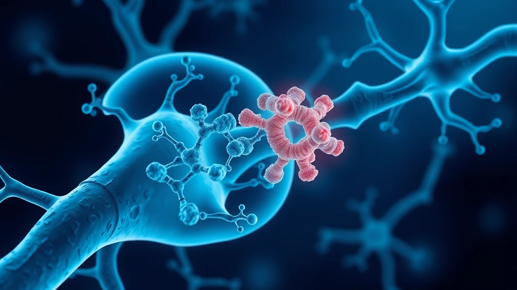
Antibodies are among medicine’s most powerful tools with a significant limitation: they normally work outside cells. Many of the most damaging proteins in brain diseases do their harm inside neurons. Researchers have used AI to bridge that gap.
A team at the University of Essex, led by Dr Caitlin O’Shea, engineered intrabodies — antibody fragments designed to function inside human cells — using AI software developed in the laboratory of Nobel laureate David Baker. The work was published in Nature Communications.
Why antibodies stop at the cell membrane
Antibodies evolved to work in blood and tissue fluid, and their structure reflects that environment.
They depend on disulphide bonds — chemical links requiring oxidising conditions to form and hold. The inside of a cell is chemically reducing, the opposite condition, so those bonds do not form properly and the antibody fails to fold correctly.
The result is that a molecule performing beautifully outside a cell becomes a misfolded, aggregating mess inside one. That is why therapeutic antibodies target surface receptors and circulating proteins, and why intracellular targets have largely been the domain of small molecules.
The charge insight
The team’s key finding is that electrical charge determines whether an antibody stays stable inside a cell or clumps together.
“Antibodies usually have the wrong charge to exist inside cells without sticking together,” O’Shea explained.
The underlying physics is intuitive once stated. Proteins carrying strong net charge repel one another and stay dispersed; proteins near neutral charge lack that repulsion and aggregate. Antibodies evolved in an environment where this was not a constraint, so their surface charge was never selected for intracellular stability.
Identifying a single physical property as the determining factor is what makes the approach scalable — charge can be calculated from sequence and modified by substituting surface residues, without redesigning the binding site.
What they built
By mapping charge properties across millions of antibodies, the team converted 672 different antibodies into functional intrabodies, and built molecules aimed at proteins implicated in Alzheimer’s, Parkinson’s, motor neurone disease and Huntington’s.
The number matters more than any individual molecule. Converting one antibody could be luck; converting 672 demonstrates a general method rather than a special case, and suggests the rule holds across diverse antibodies rather than applying to a favourable subset.
Why these diseases specifically
The four named conditions share a defining feature: each involves a protein misbehaving inside cells.
Tau accumulates within neurons in Alzheimer’s. Alpha-synuclein aggregates inside cells in Parkinson’s. TDP-43 mislocalises within neurons in motor neurone disease. Mutant huntingtin accumulates inside cells in Huntington’s.
All four have been extremely difficult to drug. Small molecules can enter cells and struggle to bind these targets, which are frequently disordered proteins without the defined pockets small molecules need. Antibodies bind such targets well and cannot get in. Intrabodies would combine the binding capability with the access.
The problem this does not solve
Making an antibody stable inside a cell is one obstacle. Getting it into the cell in the first place is another, and it is not addressed here.
Antibodies are large molecules that do not cross cell membranes, and neurons sit behind the blood-brain barrier. Delivering an intrabody to neurons in a living brain would require gene therapy encoding it so cells produce it themselves, or a delivery technology that does not currently exist at the necessary efficiency.
The gene therapy route is plausible — and inherits every difficulty of gene therapy for neurodegenerative disease, including reaching enough neurons and the permanence of the intervention.
The open-sharing commitment
The research was funded by the MND Association, and the team says it will freely share the molecules with other scientists.
That is more consequential than it sounds. 672 validated intrabodies distributed across laboratories means many groups testing many targets in parallel, rather than one group working through them sequentially.
Charity funding makes it possible: a commercial developer would have strong reasons to keep such a library proprietary, and patient organisations funding research generally prioritise the field moving over any single group’s position in it.
What this is
Early-stage research — a design toolkit and proof of concept, not a therapy. It establishes that a longstanding structural obstacle has a tractable solution and provides the reagents to explore what that enables.
What protein design has become
The role of AI here is worth characterising precisely, because it differs from how such work is usually described.
The model did not invent intrabodies or discover that charge matters. Researchers identified the physical principle; the computational tools made it possible to apply that principle across millions of antibody sequences and to redesign surface residues without destroying binding.
That is the pattern in most successful computational protein design: a human insight about biophysics, executed at a scale and precision no manual effort could reach. The tools from Baker’s laboratory, which produced the Nobel-recognised advances in this field, are essentially very good at answering “what sequence would fold into this shape with these properties” — a question that was intractable until recently.
What has changed is that redesigning a protein to have specified physical characteristics has moved from a research project to a procedure. Converting 672 antibodies would have been a career’s work by earlier methods; it is now a study.
Making the molecules openly available could accelerate work across many laboratories, which for diseases where progress has been slow is a reasonable thing to hope for. Early-stage research; not medical advice.