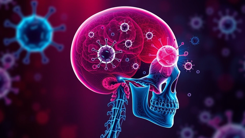
The brain has long been considered walled off from the immune system. New research reveals a line of defence hiding in plain sight: immune outposts within the skull’s bone marrow that respond to brain cancer.
Researchers at Washington University School of Medicine, led by Jonathan Kipnis with first author Jang Hyun Park, identified lymph-node-like immune structures in skull bone marrow, published in Nature.
The idea being overturned
The brain was classically described as immune-privileged — separated from immune surveillance by the blood-brain barrier and lacking the lymphatic drainage other organs use to deliver samples of their contents to lymph nodes.
That picture has eroded substantially over the past decade, most importantly with the recognition that the dura mater — the tough membrane surrounding the brain — contains lymphatic vessels after all. The brain does communicate with the immune system; the question has been how, and where the conversation happens.
What they found
The structures contain T follicular helper cells and antibody-making B cells, connected to the brain through the dura mater’s lymphatic vessels and physical channels.
That cellular composition is the important detail. T follicular helper cells and B cells together are the machinery of germinal centres — the specialised structures inside lymph nodes where antibody responses are refined and matured. Finding them organised in skull bone marrow means this is not a scattering of immune cells but a functional lymphoid tissue positioned immediately adjacent to the brain.
The physical channels matter too. Direct connections between skull marrow and the meninges mean immune cells and molecules can pass between them without entering the general circulation, which is a far more intimate arrangement than had been assumed.
Evidence they are doing something
In glioblastoma models, these skull immune hubs mounted rapid responses before distant lymph nodes detected the tumour.
That timing is the strongest evidence for their role. A structure that reacts first is receiving information first, which is what proximity and direct connection would predict.
The causal test followed: when researchers disrupted the hubs, tumours grew faster and survival fell. Removing a structure and watching outcomes worsen establishes it was contributing, rather than merely reacting.
Similar immune cells were also found in human skull bone marrow, which addresses the obvious objection that this might be a mouse peculiarity.
Why glioblastoma is the right test case
Glioblastoma is the most aggressive primary brain cancer and has proven almost entirely resistant to immunotherapy. Checkpoint inhibitors transforming outcomes in melanoma and lung cancer have achieved essentially nothing here.
The standard explanation is that glioblastoma is immunologically cold — poorly infiltrated by T cells, surrounded by suppressive signals, and in a site the immune system struggles to reach. Discovering a natural anti-tumour response already operating from the skull reframes that: the response exists and is being outpaced, which is a different problem from no response at all.
The therapeutic experiment
The team tested a gel containing three immune-boosting signals — a CD40 agonist, IL-21 and interferon-gamma — applied under the scalp. Treated mice showed better tumour rejection and longer survival.
The delivery route is the striking part. Applying a gel under the scalp places the drug against the skull, from which it can reach the marrow hubs directly — entirely sidestepping the blood-brain barrier that defeats most attempts to treat brain disease.
The chosen signals fit the biology: CD40 activation and IL-21 both support the T follicular helper and B cell responses these structures are built around, while interferon-gamma broadly enhances anti-tumour immunity.
Why the delivery route could matter more than the drug
If immune structures adjacent to the brain can be manipulated from outside the skull, that is a genuinely accessible target — a scalp application is a minor procedure compared with anything requiring intracranial access.
Kipnis said the work “fundamentally change[s] our current understanding of neuroimmunology,” with possible implications beyond cancer including Alzheimer’s and Parkinson’s. Immune involvement in both is well established, and a route to modulating brain-adjacent immunity without crossing the barrier would apply to either.
The caveats
The results are preclinical. Finding similar cells in human skull marrow establishes the structures exist in people; it does not establish they function the same way or respond to the same manipulation.
Where this fits in a decade of neuroimmunology upheaval
This finding belongs to a run of discoveries that have progressively dismantled the idea of the brain as immunologically isolated.
The sequence began with the identification of functional lymphatic vessels in the dura mater, which established a drainage route nobody had believed existed. Subsequent work described the glymphatic system clearing waste from brain tissue, and demonstrated that skull bone marrow supplies immune cells directly to the meninges rather than those cells arriving from distant bones through circulation.
Each step revealed anatomy that had been present all along in tissue examined for over a century. The structures were missed partly because standard dissection removes the dura and discards it, and partly because a strong prior belief about immune privilege shaped what people looked for.
The cumulative picture is a brain in continuous immune conversation through channels adjacent to it rather than through general circulation — which is why therapeutic access from outside the skull is a plausible idea rather than a fanciful one.
Broadly stimulating immunity adjacent to the brain also carries obvious risk — inflammation in the central nervous system is damaging, and the line between a productive anti-tumour response and harmful neuroinflammation is not one mouse survival data can fully characterise. Preclinical research; not medical advice.