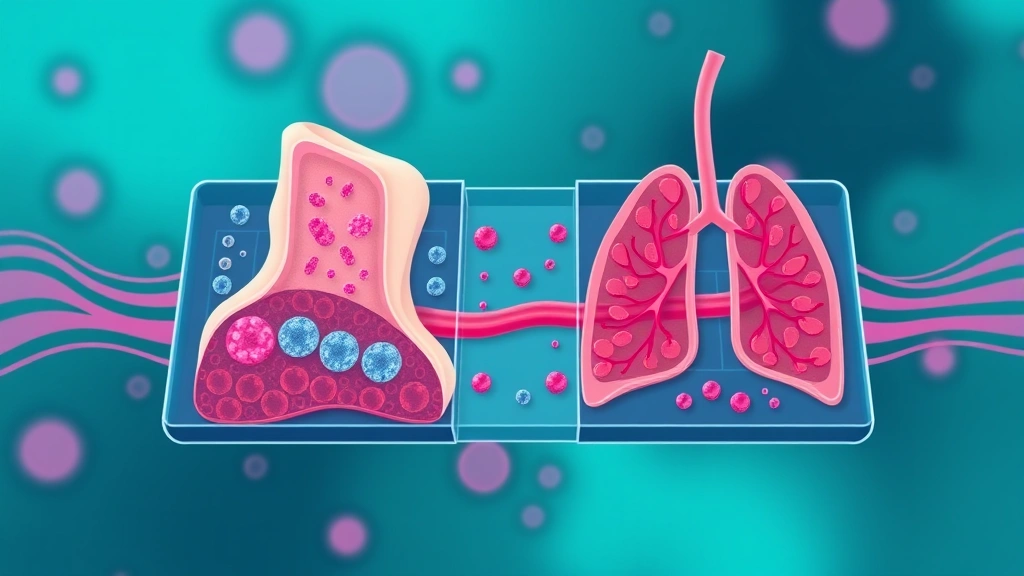
Scientists have built a miniature human body-on-a-chip that lets them watch, in real time, one of cancer’s deadliest tricks: preparing distant organs for invasion before tumour cells arrive.
Researchers at Columbia University, led by Gordana Vunjak-Novakovic, connected millimetre-sized engineered human bone and lung tissues — grown from induced pluripotent stem cells — through a vascular circulation channel. The work appeared in Science Translational Medicine.
Why metastasis is so hard to study
Metastasis, not the original tumour, causes most cancer deaths. A localised breast tumour is frequently curable; one that has spread to bone, lung, liver or brain generally is not.
Yet the process is nearly impossible to observe. In patients, metastasis is detected once deposits are large enough to image — long after the events that established them. The critical steps happen invisibly: cells entering the bloodstream, surviving circulation, exiting into tissue, and establishing themselves somewhere new.
Animal models allow observation and introduce their own problem, since mouse tissue is not human tissue and organ-specific patterns of spread differ between species.
The organ-specificity puzzle
Different cancers metastasise to characteristic sites. Breast cancer favours bone, lung, liver and brain; prostate cancer overwhelmingly targets bone. This is not explained by blood flow alone — circulating cells pass through many organs they never colonise.
The long-standing explanation holds that successful metastasis requires compatibility between the cancer cell and the tissue it lands in. Studying that compatibility requires a system containing multiple human tissues connected by circulation, which is precisely what did not exist.
What the chip does
When breast cancer cells were introduced into the flow, they showed organ-specific colonisation patterns matching human metastasis, gravitating toward bone or lung with distinct tissue-damage signatures.
Reproducing organ preference is the validation that matters. A system where cancer cells settled randomly would model circulation without modelling metastasis; recapitulating the pattern seen in patients suggests the mechanisms determining that pattern are operating in the device.
A selectively permeable barrier of endothelial cells separated the tissues from the blood channel, letting researchers observe cancer cells adhere to, cross, and settle into tissue.
That barrier is essential to the design. Crossing the vessel wall is a discrete, difficult step in metastasis, and a system without a realistic barrier would let cells into tissue too easily — skipping the stage where most circulating cancer cells fail.
The pre-metastatic niche
The chip captured how cancer cells condition an organ before colonising it — pre-metastatic niche formation.
This is one of the more unsettling ideas in cancer biology. A primary tumour releases signalling molecules and vesicles into circulation that alter distant tissue before any cancer cell arrives — recruiting particular immune cells, changing the local matrix, making the site receptive.
Metastasis on this account is not opportunistic. The ground is prepared in advance, remotely, by a tumour that has not yet spread.
“Cancer is very smart, unfortunately. We learned how the cells cross barriers to get from blood circulation into the tissues,” Vunjak-Novakovic said.
Why using stem-cell-derived human tissue matters
Growing the bone and lung tissues from induced pluripotent stem cells means the system is entirely human, and in principle can be built from a specific individual’s cells.
That opens a possibility conventional models cannot offer: constructing a chip from a patient’s own tissue and their own tumour, and testing how that combination behaves. Whether that is practical at scale is a separate question, but the architecture allows it.
What it could be used for
The obvious application is drug screening against metastasis specifically, rather than against tumour growth.
Almost all cancer drug development measures whether a compound shrinks a tumour. Very little measures whether it prevents spread, largely because there has been no tractable way to test that. A platform where the steps of metastasis are observable is a platform where interventions blocking each step can be evaluated.
Preventing niche formation is a particularly interesting target, since it would mean intervening before metastasis rather than treating it after.
The limits
A two-organ chip is a substantial simplification of a body. It has no immune system in any complete sense, no liver metabolising drugs, no nervous system, and millimetre-sized tissues rather than organs.
Timescales differ too — metastasis in patients unfolds over months to years, and a device cannot be maintained that long.
Why bone and lung were the right pair to build
The choice of tissues is not arbitrary, and it reflects where breast cancer actually goes.
Bone is the most common site of breast cancer metastasis, and bone metastases cause much of the disease’s morbidity — fractures, spinal cord compression, severe pain and dangerous elevations in blood calcium. They are also where cancer cells are thought to lie dormant for years, which connects directly to the problem of late recurrence.
Lung is the other major site and behaves differently, with metastases there tending to grow rather than lie dormant.
Building a chip with both means the system can be interrogated about why the same circulating cells behave differently depending on where they land — dormancy in one tissue, proliferation in another. That comparison is impossible in a single-tissue model and difficult in a patient, and it is arguably the most valuable question the platform is positioned to ask.
What it offers is not a replacement for animal models or patients but a system where specific mechanistic questions about human cells crossing human barriers can be asked directly and watched happening. Research news, not medical advice.